By Dr. Blayne Mozisek, MS, DVM, MAM, DACPV
Check Your Flock for Intestinal Parasites at Home
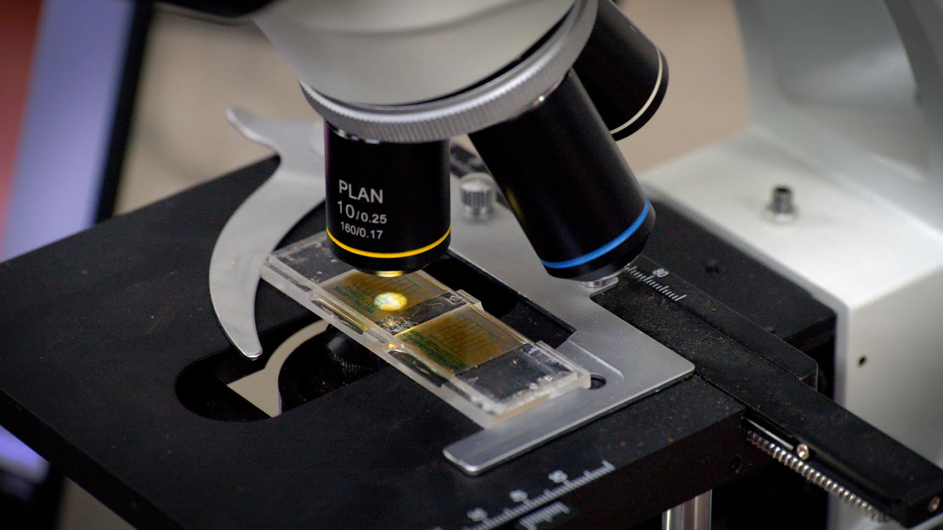
A fecal flotation is a simple way to look for parasite eggs and coccidia oocysts in your chickens’ droppings using a microscope.
This guide covers two quantitative flotation protocols that backyard poultry keepers can perform at home without laboratory centrifuges or specialized equipment: a modified Wisconsin passive sugar flotation, and a McMaster counting-chamber technique. Both use the same specific gravity (SG) 1.27 sugar flotation solution.
Common parasites that may be detected include:
- Eimeria spp. (coccidia)
- Ascaridia galli (large roundworm)
- Heterakis gallinarum (cecal worm)
- Capillaria spp. (capillary/thread worms)
- Some cestode (tapeworm) eggs or egg packets
- Other less common gastrointestinal parasite eggs
Important: A fecal flotation can tell you whether parasite eggs or oocysts are present and provide an estimate of how many are being shed. It does not, by itself, determine whether those parasites are causing disease or whether treatment is necessary.
Choosing Your Method: Wisconsin vs. McMaster
Both protocols provide standardized parasite-count estimates, but they do so differently. McMaster is a quantitative counting-chamber method that reports estimated eggs per gram (EPG) or oocysts per gram (OPG). The passive Wisconsin adaptation provides a whole-coverslip count that can be expressed per gram, but it has not been validated as a laboratory-equivalent quantitative method. Read the comparison, then choose the method that best matches what you’re trying to accomplish.
Side-by-Side Comparison
| Factor | Modified Wisconsin (Passive) | McMaster |
|---|---|---|
| Sample mass | 3.0 g feces | 4.0 g feces |
| Flotation solution | 10 mL SG 1.27 sugar solution | 60 mL SG 1.27 sugar solution |
| Chamber | Standard microscope slide + coverslip | Two-chamber McMaster counting slide |
| Flotation time | 20 minutes (passive) | Fill chambers, rest 3–5 minutes, then examine |
| Reading area | Entire coverslip | Two counting grids |
| Math | Total count ÷ 3 = EPG/OPG | Combined grid count × 50 = EPG/OPG |
| Time per sample | ~25 minutes | ~10 minutes |
| Detection limit | No validated at-home detection limit; whole-coverslip scan may improve low-count detection | Approximately 50 EPG with the 4 g/60 mL final-suspension, two-grid protocol described here |
| Best for | Sick bird workup, ruling parasites in/out, Capillaria screening | Routine monthly/quarterly flock trending across many birds |
Modified Wisconsin, Pros and Cons
Pros:
- Greater low-count detection potential. Because the entire coverslip is scanned, this passive whole-coverslip method may detect parasite stages that a limited-volume McMaster count misses. A specific validated detection limit has not been established for this at-home passive adaptation.
- Useful when Capillaria is a concern. Capillaria eggs may be shed in low numbers, so examining the entire coverslip can improve the chance of finding eggs that could fall below the McMaster counting threshold.
- No specialized slide required. Uses ordinary microscope slides and coverslips.
- Simple arithmetic. Total count divided by 3.
- Ideal when the question is “is this bird carrying parasites at all?”, clinical workup, pre-purchase screening, or before a treatment decision.
Cons:
- Slower. Twenty minutes of settling plus a full-coverslip scan means about 25 minutes per sample.
- Not built for high throughput. If you’re monitoring several groups of birds monthly, the time adds up quickly.
- Passive technique. Less sensitive than a properly centrifuged Wisconsin performed in a diagnostic lab.
McMaster, Pros and Cons
Pros:
- Fast. About 10 minutes per sample once the technique is practiced.
- Highly repeatable for trending. The counting-chamber geometry standardizes the volume you look at, which makes flock-level monthly or quarterly comparisons very consistent.
- Ideal for routine flock monitoring. If you have several coops, breeds, or age groups and you want a running record of parasite pressure over time, this is the method built for that job.
- Well-established quantitative method. McMaster counting chambers are widely used in veterinary parasitology to estimate fecal egg counts, particularly when repeatable counts are needed for monitoring over time.
Cons:
- Higher detection limit than whole-coverslip flotation. With the 4 g/60 mL final-suspension protocol and a standard two-grid McMaster slide described here, one egg counted corresponds to approximately 50 EPG. Low-level infections can therefore still be missed.
- Requires a McMaster slide. These are inexpensive but must be purchased; they cannot be substituted with a regular coverslip.
- Less sensitive for Capillaria and tapeworms. Both shed relatively few eggs; light infections can slip below the McMaster detection threshold.
- Sample must be remixed before filling each chamber. Eggs redistribute rapidly, and forgetting to remix biases the count.
Quick Decision Framework
- A bird looks sick or off, and you want the most sensitive at-home test → Wisconsin.
- You suspect Capillaria specifically → Wisconsin.
- You’re doing routine monthly or quarterly flock trending → McMaster.
- You’re comparing several groups against each other → McMaster (or run Wisconsin on all of them with the same 3 g standard mass, either works if you’re consistent).
- You want to run one baseline right now and see what’s out there → Either method is fine. Wisconsin is more forgiving if you’re new to microscopy; McMaster is faster once you have the slide.
Whichever method you choose, stick with it for any trend comparison. Wisconsin EPG values and McMaster EPG values are not directly interchangeable because their detection thresholds differ.
What You’ll Need
Shared Equipment (Both Methods)
- Compound microscope with 10× and 40× objectives
- Microscope camera or phone adapter (optional, but strongly recommended for photographing parasites for identification)
- Hydrometer capable of measuring approximately SG 1.20 to 1.30
- Digital kitchen or laboratory scale accurate to at least 0.1 g
- Small disposable cup
- Small mixing stick, craft stick, or disposable spoon
- Small funnel
- Tea strainer, fine kitchen strainer, or two layers of cheesecloth
- Disposable transfer pipette or medicine dropper
- Measuring cup
- Timer
- Disposable gloves
Wisconsin-Specific
- Microscope slides
- Coverslips
- Small straight-sided tube or vial approximately 15 to 20 mL in volume
McMaster-Specific
- Two-chamber McMaster counting slide (Whitlock, universal, or equivalent)
- Larger mixing container, approximately 100 mL capacity
For the Flotation Solution (Both Methods)
- Ordinary white granulated table sugar
- Water
- Hydrometer
- Clean container with a lid
Part 1: Make Your Sugar Flotation Solution (Both Methods)
The sugar solution needs to be dense enough for parasite eggs and oocysts to float to the surface.
For this test, your target is:
Specific Gravity = 1.27
The hydrometer is important because measuring sugar and water alone cannot guarantee the final specific gravity. Temperature, evaporation, measurement differences, and incomplete mixing can all affect the final solution.
Starting Recipe
Combine 1 lb (454 g) white granulated sugar with 12 fl oz (355 mL) water.
Directions
- Place the water in a saucepan or heat-safe container.
- Gradually add the sugar while stirring.
- Warm the mixture gently while continuing to stir until the sugar is completely dissolved.
- Do not boil the solution. Excessive heating can evaporate water and change the specific gravity.
- Remove the solution from the heat.
- Allow it to cool completely to room temperature.
- Pour enough solution into a container that allows your hydrometer to float freely.
- Insert the hydrometer and read the specific gravity at the liquid surface.
Your Target: SG 1.27
A reading close to 1.27 is what you want.
If the SG Is Too Low
The solution contains too much water relative to sugar.
Gently reheat the solution and add a small amount of sugar (approximately 1 tablespoon at a time), stirring until it is completely dissolved. Alternatively, you can gently heat the solution to evaporate a small amount of water and increase its concentration. If using evaporation, heat gradually and monitor closely; do not allow vigorous boiling or scorching. In either case, allow the solution to cool completely to room temperature before checking the SG again. Repeat as needed until the SG reaches approximately 1.27.
If the SG Is Too High
The solution is too concentrated.
Add approximately 1 teaspoon of water at a time, mix thoroughly, and check the SG again. Repeat until the SG reaches approximately 1.27.
Always check specific gravity at room temperature. Temperature affects the density of the solution and can change the hydrometer reading.
Once prepared, place the solution in a clean, tightly closed container and label it:
Fecal Flotation Solution, SG 1.27, Not for Consumption
If the solution develops mold, fermentation, unusual cloudiness, or contamination, discard it and make a fresh batch.
Part 2: Collect Your Sample (Both Methods)
Fresh samples provide the best results.
Testing One Chicken
Collect a fresh dropping directly after the bird defecates or from a clean surface where you are confident it came from that bird. Avoid collecting excessive bedding, dirt, feathers, or other material with the dropping.
Testing Your Flock
For routine flock monitoring, you can create a pooled sample.
Collect several fresh droppings from different locations beneath the roost or from several birds in the same flock or management group. Combine them and mix them thoroughly before taking your test sample.
Do not combine samples from groups you specifically want to compare, for example chicks and adult hens, sick and healthy birds, or birds housed in different pens. Keep those samples separate.
If You Can’t Test It Immediately
Place the sample in a sealed container or plastic bag and refrigerate it. Do not freeze it. Test the sample as soon as practical.
Modified Wisconsin Protocol
The passive sugar flotation described below uses 3.0 g of feces in 10 mL of SG 1.27 solution and a 20-minute passive settling time. Follow this section end-to-end if you selected the Wisconsin method.
Part 3W: Prepare the Sample
Put on disposable gloves before handling feces.
- Homogenize the feces before weighing your test portion. If the sample was collected in a sealable plastic bag, keep the bag closed and knead the droppings thoroughly through the outside of the bag until the sample is as uniform as practical. This is especially important for pooled samples.
- Weigh exactly 3.0 grams. Place your disposable cup on the scale and tare or zero the scale. Add feces until you have 3.0 g. Using the same sample weight every time makes your results much easier to compare.
- Measure 10 mL of sugar solution. Add 10 mL of SG 1.27 sugar flotation solution to the 3 g fecal sample.
- Mix thoroughly. Use a craft stick, spoon, or similar disposable tool to mash and stir the feces into the solution. Break apart clumps. You want the fecal material suspended throughout the liquid rather than sitting in large pieces at the bottom.
- Strain the mixture. Place a small funnel over your tube. Place a fine tea strainer or two layers of cheesecloth inside the funnel. Pour the fecal mixture through the strainer and into the tube. Gently press the remaining material to recover liquid, but don’t force large pieces of debris through the strainer.
Part 4W: Float the Parasites
- Fill the tube completely. Using a transfer pipette or dropper, add additional SG 1.27 sugar solution until the liquid reaches the very top of the tube. Continue adding solution carefully until the surface forms a slight rounded dome, or convex meniscus, above the rim. Think of it as a tiny bubble of liquid sitting just above the tube.
- Add the coverslip. Carefully place a clean coverslip directly on top of the liquid. The coverslip should contact the liquid. Try not to trap a large air bubble underneath it.
- Wait 20 minutes. Leave the tube completely undisturbed with the coverslip in place for 20 minutes. During this time, parasite eggs and oocysts that are less dense than the SG 1.27 sugar solution can rise through the liquid and collect against the underside of the coverslip. Don’t bump or move the tube during this period.
- Remove the coverslip. After 20 minutes, lift the coverslip straight upward. Don’t slide it across the rim of the tube.
- Place it on a microscope slide. Immediately place the coverslip wet side down onto a clean microscope slide. Your sample is ready to examine.
Part 5W: Examine the Slide
Start with your microscope’s 10× objective. With a standard 10× eyepiece, this gives approximately 100× total magnification. This is the primary magnification you will use to search for parasite eggs and coccidia oocysts.
Scan the entire coverslip. Start in one corner. Move across the slide in a straight line. When you reach the opposite edge, move down slightly and scan back in the other direction. Continue this back-and-forth pattern until you’ve examined the entire coverslip.
Think of it like mowing a lawn: you want your paths to overlap slightly so you don’t miss an area.
When you find something that may be a parasite, center it in your field of view. Switch to the 40× objective (approximately 400× total magnification) when you need a closer look at its morphology. Return to 100× to continue scanning.
Part 6W: Count and Calculate, Wisconsin Math
Keep a separate count for each parasite category while scanning the entire coverslip.
Because you started with 3.0 grams of feces, divide your total count for each parasite category by 3.
EPG or OPG = Total Count ÷ 3
Example: 30 Ascaridia/Heterakis-type eggs across the coverslip → 30 ÷ 3 = 10 EPG.
Note: Ascaridia galli and Heterakis gallinarum eggs are very similar and are generally not reliably distinguished from one another during a routine backyard fecal examination. Record them as Ascaridia/Heterakis-type eggs unless identification is supported by additional expertise or testing.
McMaster Protocol
The McMaster technique described here uses 4.0 g of feces and enough SG 1.27 sugar flotation solution to produce 60 mL of final suspension, then examines two standard McMaster counting grids. This increases analytical sensitivity to approximately 50 EPG with a standard two-grid slide. Follow this section end-to-end if you selected the McMaster method.
Part 3M: Prepare the Sample
Put on disposable gloves before handling feces.
- Homogenize the feces before weighing your test portion. If the sample was collected in a sealable plastic bag, keep the bag closed and knead the droppings thoroughly through the outside of the bag until the sample is as uniform as practical. This is especially important for pooled samples.
- Weigh exactly 4.0 grams. Place your mixing container on the scale and tare or zero it. Add feces until you have 4.0 g.
- Prepare a final suspension volume of 60 mL. Add SG 1.27 sugar flotation solution to the 4.0 g fecal sample, mix thoroughly, then add additional flotation solution as needed so the total final suspension volume is 60 mL. Keeping the final volume standardized is what makes the ×50 McMaster multiplier work with two standard 0.15-mL grids.
- Mix thoroughly. Mash and stir the feces into the solution until the fecal material is fully suspended and clumps are broken apart.
- Strain the mixture. Pour through a tea strainer or two layers of cheesecloth into a second clean container. This removes coarse debris that would obstruct the counting chambers.
Part 4M: Fill the Counting Chambers
The McMaster slide has two counting chambers, each with an etched grid on its underside.
- Remix the strained suspension. This is the most commonly skipped step and it will bias your count. Stir or gently swirl the suspension for a few seconds immediately before filling each chamber. Eggs settle and rise unevenly within the container over even short periods.
- Fill the first chamber. Draw suspension into a transfer pipette immediately after mixing, and fill the first chamber completely. There should be no air bubbles and no dry spots under the etched grid.
- Remix again, then fill the second chamber.
- Rest the slide flat for 3 to 5 minutes. Parasite eggs and oocysts will rise to the underside of the coverglass (the grid surface), while heavier debris settles to the bottom of the chamber.
Part 5M: Examine and Count
Place the slide on the microscope stage with the counting grids facing up.
Use the 10× objective (approximately 100× total magnification) and focus on the etched grid lines. This is the plane where floated eggs and oocysts accumulate.
Count only within the grid area of each chamber. Do not count eggs that fall outside the grid boundary. Include eggs touching the top or left grid line; exclude eggs touching the bottom or right grid line (a standard microscopy convention that prevents double-counting).
Keep a separate count for each parasite category.
Part 6M: Count and Calculate, McMaster Math
Combine the counts from both chambers, then multiply by 50 to get EPG or OPG.
EPG or OPG = (Chamber 1 count + Chamber 2 count) × 50
Example: 3 Ascaridia/Heterakis-type eggs in Chamber 1 + 2 in Chamber 2 = 5 total → 5 × 50 = 250 EPG.
The multiplier of 50 comes from examining 0.3 mL total across two standard 0.15-mL grids from a 60 mL final suspension prepared with 4.0 g of feces. If you use a McMaster slide with different chamber or grid volumes, calculate the appropriate multiplier for that slide or follow the manufacturer’s validated protocol.
What Are You Looking For? (Both Methods)
Eimeria spp. (Coccidia)
Look for small oval to round oocysts. Their size and appearance vary among Eimeria species.
Finding Eimeria oocysts does not automatically mean the chicken has clinical coccidiosis. Healthy chickens can shed oocysts.
Ascaridia / Heterakis-Type Eggs
These are generally larger, oval, thick-walled eggs.
Eggs of Ascaridia galli and Heterakis gallinarum can look similar under routine microscopy. Backyard testing should generally record them as Ascaridia/Heterakis-type unless you have sufficient experience and morphology to confidently distinguish them.
Capillaria spp.
Look for characteristic elongated or barrel-shaped eggs with bipolar plugs (small plug-like structures at each end).
Capillaria is easier to miss with McMaster because it sheds relatively few eggs. If you strongly suspect Capillaria, run the Wisconsin protocol instead — or in addition.
Tapeworms
Tapeworm eggs or egg packets may occasionally be seen.
Tapeworm infections can be particularly difficult to detect by fecal flotation because eggs may not be distributed evenly through the feces and some species shed segments containing eggs intermittently. These proglottids (tapeworm sections) are most often seen grossly on the top of fresh, formed droppings.
A negative fecal flotation does not rule out tapeworm infection regardless of which method you used.
Understanding Your Results
A fecal flotation is an important tool, but parasite numbers should never be interpreted by themselves.
The number of eggs or oocysts shed can be affected by:
- Parasite species
- Number and maturity of parasites
- Age of the chicken
- Immunity
- Recent treatment
- Time of sample collection
- Distribution of eggs within the feces
- Sample storage and handling
- Quality of the flotation technique
This is particularly important with coccidia.
Finding Eimeria oocysts does not equal a diagnosis of coccidiosis.
Most chickens are exposed to Eimeria, and healthy birds may shed oocysts as immunity develops. OPG should be considered alongside the bird’s age, clinical signs, flock history, lesions when available, and overall performance.
Similarly, a negative result does not prove that your flock is parasite-free, especially with the McMaster protocol described here, which has a calculated detection limit of approximately 50 EPG when both standard grids are counted.
Comparing Wisconsin and McMaster Results
Do not directly compare EPG values across methods. A count of 100 EPG on Wisconsin is not the same as 100 EPG on McMaster because their detection thresholds and sampling geometries differ. For any trend you want to follow over time, stay with one method.
If you want to switch methods, run both methods on the same sample for a few rounds to establish the offset for your specific setup before treating the new method as your baseline.
When There Are Too Many to Count
Occasionally a slide may contain so many eggs or oocysts that accurately counting isn’t practical.
Record the result as TNTC (Too Numerous to Count). Do not estimate a number.
If an accurate quantitative result is needed, the sample should be repeated using an appropriately standardized dilution method. McMaster with a higher dilution or a chamber-counted subsample is the standard next step. When a sample contains parasite eggs or oocysts that are TNTC, this should be considered a result that requires intervention.
Record Your Results
Flock/Bird: __________________________
Date: _______________________________
Age: ________________________________
Method used: ☐ Wisconsin (3 g / 10 mL / ÷3) ☐ McMaster (4 g / 60 mL final / ×50)
Individual or pooled sample: __________________
Clinical signs: _______________________________
Recent dewormer/anticoccidial use: ______________
Flotation solution SG: _________________________
Flotation time: _______________________________
| Parasite | Raw Count | Calculation | Result |
|---|---|---|---|
| Ascaridia/Heterakis-type | ÷ 3 or × 50 | EPG | |
| Capillaria | ÷ 3 or × 50 | EPG | |
| Other helminths | ÷ 3 or × 50 | EPG | |
| Eimeria | ÷ 3 or × 50 | OPG |
Notes: _______________________________________
Tips for More Reliable Results
- Use fresh feces whenever possible.
- Verify that your sugar solution is SG 1.27 every batch.
- Use the same sample mass every time within a method (3.0 g for Wisconsin, 4.0 g for McMaster).
- Thoroughly mix pooled samples before removing the test portion.
- Break apart fecal material completely in the flotation solution.
- McMaster only: remix the strained suspension immediately before filling each chamber.
- Wisconsin only: keep the flotation time standardized at 20 minutes and scan the entire coverslip.
- Use the same scanning pattern every time.
- Photograph questionable findings through your microscope.
- Keep samples from different age groups or management groups separate when comparison matters.
- Record recent parasite treatments with every result.
- Do not mix methods within a single trending series.
Common Mistakes
My hydrometer reads below 1.27.
Your solution is too dilute. Add more sugar, dissolve it completely, cool to room temperature, and retest.
My hydrometer reads above 1.27.
Add a small amount of water, mix thoroughly, and retest.
(Wisconsin) I can’t get a rounded meniscus.
Your tube may be too wide or isn’t completely full. Add flotation solution one drop at a time until the liquid rises slightly above the rim.
(McMaster) My chambers have air bubbles or dry spots.
Refill immediately. Air pockets under the grid cause both false negatives (eggs missed) and biased counts. Draw fresh suspension after remixing and refill in one smooth motion.
(McMaster) My two chamber counts are very different from each other.
You probably didn’t remix the strained suspension before filling the second chamber. Eggs redistribute rapidly. Always remix. If chambers still disagree by more than roughly 30 to 50 percent after remixing, prepare a fresh sample.
There is a lot of debris under the microscope.
Some debris is expected. Make sure the sample was strained adequately and avoid forcing coarse fecal material through the strainer.
I see bubbles everywhere.
Air bubbles are extremely common and can initially look suspicious. Adjust the microscope’s focus up and down. Bubbles usually have a highly refractile circular border and lack the internal structures expected in parasite eggs or oocysts.
I didn’t find anything.
A negative test means no parasite stages were detected in the sample using this method. It does not prove that the bird or flock is parasite-free, particularly with McMaster, which has a higher detection floor.
Safety
Poultry feces can contain organisms capable of causing disease in people.
- Wear disposable gloves.
- Keep fecal-testing equipment away from food-preparation areas.
- Do not use kitchen equipment for food again after dedicating it to fecal testing.
- Wash your hands thoroughly afterward.
- Clean and disinfect the work surface when finished.
- Keep prepared flotation solution labeled and away from children and animals.
Important Limitations of At-Home Methods
Both protocols avoid centrifugation so they can be performed practically by backyard poultry owners. The Wisconsin adaptation uses standing (passive) flotation under a coverslip; the McMaster method uses flotation within a calibrated counting chamber.
Passive flotation is less sensitive than properly performed centrifugal flotation. Low-level infections may be missed by either method, and are more likely to be missed by McMaster than by Wisconsin.
Results should be reported as Passive sugar fecal flotation (Wisconsin, SG 1.27) or Passive McMaster (SG 1.27) so future readers of your records know exactly what technique produced the numbers.
If your bird is sick, losing weight, producing abnormal droppings, has blood in the feces, or you continue to suspect parasites despite a negative test, veterinary evaluation and/or professional diagnostic testing is recommended.
TPD Marketplace: Build Your At-Home Fecal Testing Kit
Everything needed to perform either test can be assembled into a simple home microscopy kit:
- Compound microscope with 10× and 40× objectives
- Microscope camera / phone adapter
- Specific-gravity hydrometer (SG 1.20–1.30)
- Digital scale (0.1 g accuracy)
- Microscope slides
- Coverslips
- Two-chamber McMaster counting slide (for the McMaster protocol)
- Transfer pipettes
- Sample tubes
- Mail-in Fecal Exam
Need help identifying what you found? Photograph your findings through your microscope and use The Poultry Doc’s diagnostic resources or contact our team for assistance.
Scientific Basis
These protocols are adapted from established veterinary fecal-flotation and fecal egg-count techniques for practical use without a centrifuge. McMaster is a widely used quantitative fecal egg-count method. The Wisconsin-style protocol presented here is an at-home passive adaptation of a method commonly performed with centrifugation in laboratory settings. Published veterinary references support sucrose/Sheather’s sugar flotation solution at approximately SG 1.27 for recovery of many common gastrointestinal parasite eggs and coccidian oocysts.
Both home versions standardize fecal mass, flotation-solution SG, timing, and reporting to improve repeatability. The passive Wisconsin adaptation should not be treated as equivalent to a centrifuged Wisconsin laboratory result. McMaster counts are calculated from a defined chamber volume, but results still depend on sample preparation, flotation solution, chamber design, and counting technique. Because the two methods use different sampling geometries and have different analytical sensitivity, do not directly compare Wisconsin-style whole-coverslip counts with McMaster EPG/OPG values. For trend monitoring, use the same method consistently.